


Dr. Reem Alramli, DMD
What Are Dental X-Rays?
Dr. Reem Alramli answers the questions patients ask most about dental X-rays.
What is a dental X-ray?
A dental X-ray is a picture that helps us see your teeth, roots and jawbone, including the areas we cannot see during a regular exam.
When do you take dental X-rays?
We take X-rays during dental checkups, and when needed, for example if a patient has pain, swelling or other dental concerns.
Why are dental X-rays important?
Dental X-rays help us find dental problems early, before they become more serious and complicated, and allow us to plan the right treatment.
What types of dental X-rays are there?
We have different types. Bitewings help us detect cavities in between the teeth. Periapical X-rays help us see the entire tooth and the roots. Panoramic X-rays help us see the full mouth. And we have three-dimensional X-rays, called CBCT, which give us more information about bone density and other areas like the sinus and the wisdom teeth.
Are dental X-rays safe?
Yes. Dental X-rays use a very low amount of radiation, especially with modern technology, and we only take them when necessary.
What can dental X-rays show?
They can show cavities, infection, bone loss, impacted teeth and other problems that may not be visible during the exam.
Can X-rays detect gum disease?
Yes. X-rays help us identify bone loss around the teeth, which can be a sign of gum disease.
Are dental X-rays covered by insurance?
Most dental X-rays are covered by insurance for routine checkups, but the coverage depends on your specific plan.
How often should I see the dentist?
Most patients benefit from a dental exam every six months, but some may need more frequent visits depending on their gum health. Contact Harmony Cosmetic Dentistry & Implants at (832) 464-4454. We are here to help you restore your smile.
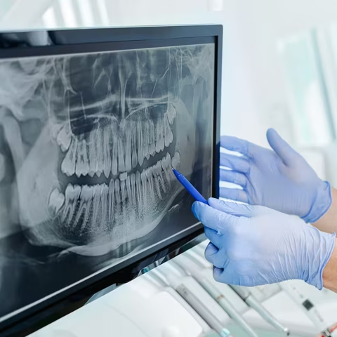
Digital X-Rays: A Closer Look at Your Oral Health
Some dental concerns hide below the surface, in places a visual exam alone cannot reach. Decay between teeth, bone loss around the roots, and issues beneath the gum line often go unnoticed until they are further along.
As part of our general dentistry services, we use digital X-ray imaging to see what a standard exam cannot show on its own. This technology gives our team a clearer, more complete view of your teeth, roots, and supporting bone, which helps guide accurate diagnosis and treatment planning from the very first visit.
What Are Digital X-Rays?
Digital X-rays capture images of your teeth and jaw using a small sensor instead of traditional film. The image appears on a computer screen within seconds, allowing our team to review it right away rather than waiting for film to develop.
This technology is used across both sides of our practice, supporting routine general dentistry evaluations as well as more detailed imaging, such as CBCT scans, for implant planning and periodontal treatment.
Benefits of Digital X-Ray Imaging
-
Earlier Detection of Hidden Issues
Digital X-rays reveal decay between teeth, bone changes, and other concerns that are not visible during a standard visual exam. Catching these issues early often means simpler, less invasive treatment down the road.
-
Lower Radiation Exposure
Digital sensors require significantly less radiation than traditional film to produce a clear, usable image. This makes the process gentler on your body while still providing the detail needed for an accurate diagnosis.
-
Faster, More Convenient Visits
Images appear on screen within seconds instead of requiring time to develop, so Dr. Reem can review your results during the same appointment. This means less waiting and quicker answers about your treatment options.
What to Expect During Your X-Ray
Taking digital X-rays is a quick, straightforward part of your visit. Here is what generally happens during this step of your appointment.
-
Step 1:
Positioning the Sensor
A small digital sensor is carefully placed in your mouth to capture the specific area our team needs to evaluate.
-
Step 2:
Capturing & Reviewing the Image
The X-ray is taken in seconds using a low dose of radiation, and the image appears on screen almost immediately for Dr. Reem to review with you during the same visit.
-
Step 3:
Identifying Any Concerns
Our team looks for decay between teeth, bone changes, and other issues that would not be visible during a standard visual exam alone.
-
Step 4:
Supporting Your Treatment Plan
These images become part of your dental record and guide any recommended treatment, while also building a history that helps us track changes in your oral health over time.
See What Digital X-Rays Can Reveal
Digital imaging has become a standard part of modern dental care because it helps our team catch problems earlier and plan treatment more accurately.
Whether you are coming in for a routine comprehensive exam or a more involved evaluation, digital X-rays help our team build a complete picture of your oral health. You can contact our office to schedule your visit and learn more about how this imaging supports your care.
Ready to schedule your digital X-ray appointment?
Our Magnolia office is accepting new adult patients. Call us today or book online.
Request An AppointmentFrequently Asked Questions
Are dental X-rays safe?
Yes. Digital X-rays use significantly less radiation than traditional film X-rays — up to 80 percent less — and the exposure is brief and highly targeted. At Harmony Cosmetic Dentistry and Implants, we take only the images necessary based on your individual health history and risk factors, and we use protective equipment as needed. The diagnostic benefit of detecting problems early far outweighs the minimal radiation involved.
How often do I need dental X-rays?
Frequency depends on your individual oral health. New patients typically receive a full set of X-rays so we can establish a complete baseline. After that, most patients with stable oral health need bitewing X-rays once a year and a full set every three to five years. Patients with a history of decay, gum disease, or complex dental work may need them more frequently. Dr. Reem will determine what is appropriate for you.
What can digital X-rays detect that a visual exam cannot?
Digital X-rays reveal decay between teeth and under existing restorations, bone loss from gum disease, infections or abscesses at the tooth roots, impacted teeth, cysts, and early signs of other structural problems — none of which are visible during a standard visual exam. They are an essential part of a complete diagnostic picture.
Do dental X-rays hurt?
No. Digital X-rays are completely non-invasive. A small sensor or film is positioned inside your mouth, you hold still for a moment, and the image is captured. There is no discomfort during the imaging itself. Some patients find the sensor placement slightly awkward, but there is no pain involved.
Will X-rays be taken at every visit?
Not necessarily. Routine checkup visits typically do not require a full set of images each time. Bitewing X-rays are usually taken once a year to check for decay between teeth, while more comprehensive imaging is scheduled less frequently. If you come in for a specific concern — pain, swelling, or a new symptom — targeted X-rays may be taken at that visit regardless of when your last set was taken.
Can I see my X-rays during my appointment?
Yes. Because our X-rays are digital, the images appear on screen almost instantly and can be enlarged for a detailed view. Dr. Reem will walk you through what she sees and explain any findings in plain language so you have a clear understanding of your oral health before any treatment is discussed.